MS imaging makes ingredients, additives and contaminants of food visible
An analytical method without dyes
mass spectrometry imaging (MS imaging) provides highly precise information on the spatial distribution of substances in many areas. Researchers at the University of Bayreuth now present exemplary new applications in food analysis in the journal "Food Chemistry". For the first time, they have succeeded in making visible an additive in dairy products and a production-related contamination in baked goods. Special ingredients that influence food quality can be detected in fruit, vegetables and meat products.The study, which was conducted in cooperation with the Bavarian Health and food safety Authority (LGL), shows the great potential of this method, not least in terms of consumer protection.
Oliver Wittek
Natamycin in cheese
To protect cheese wheels or smoked sausages from mold infestation, the surfaces are often treated with the fungicide natamycin. An EU regulation sets a limit of one milligram per square decimeter for this and also stipulates that natamycin must not penetrate deeper than five millimeters into a treated cheese wheel. However, this penetration depth cannot be described in detail using the food analysis methods commonly used to date. However, the Bayreuth research team led by Prof. Dr. Andreas Römpp has been able to use MS imaging to show for the first time where and in what quantities the fungicide occurs in different types of Gouda. The penetration of the natamycin molecules can be tracked from the rind to the inside of the cheese wheel. The scientists collaborated with the Bavarian Health and Food Safety Authority (LGL) in these investigations. Based on the results obtained, they have developed methodological standards for the identification of natamycin in cheese. "Building on this newly developed MS imaging approach, it may be possible to reduce consumer exposure to preservatives in the future," says Prof. Römpp, who is the Chair of Bioanalytic Sciences and Food Analysis at the University of Bayreuth.
Acrylamide in gingerbread
An EU regulation also sets limits for the presence of acrylamide in food. It is a cancer-promoting substance that is formed from sugar and asparagine - an amino acid - at low humidity and temperatures above 120 degrees Celsius. A method developed in Bayreuth, Germany, based on MS imaging, visualizes acrylamide distribution in traditional German gingerbread. "To do this, we had to cool the gingerbread samples to less than minus 60 degrees Celsius and then use an electric microsaw to produce gingerbread slices of two millimeter thickness. This was the only way we could detect very small amounts of acrylamide," reports Prof. Römpp.
Studies of veal sausages
The new study also shows that MS imaging is equally suitable for analyses of processed meat products. In veal sausages, water-soluble and fat-soluble components become visible, so that low-fat and high-fat regions can be clearly distinguished. Likewise, it becomes visible where substances of plant origin are found that come from admixed herbs. "However, MS imaging not only enables the localization of ingredients in meat products, but also helps, for example, in investigations of 'sticky meat' or so-called hydrolysate additives, which are supposed to feign higher quality when they are not declared on the packaging. It could therefore be useful in detecting consumer deception in meat products and better protect consumers in this respect as well," says Prof. Römpp.
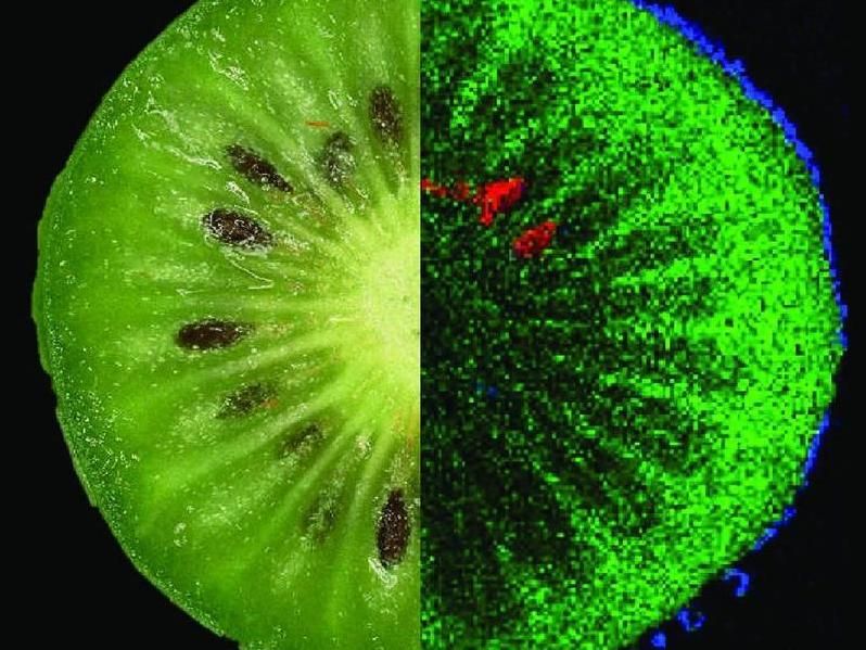
Kiwifruit and carrots
The application potential in the field of fruits and vegetables is demonstrated by studies on kiwifruit and carrots. The "mini kiwi" (Actinidia arguta) is not only sweet, but also has numerous health-promoting bioactive ingredients. Using sample slices that were only a few hundredths of a millimeter thick and cooled down to a temperature of minus 40 degrees, the Bayreuth bioanalysts visualized the distribution of several substances in the skin and flesh: sugar molecules (disaccharides), antioxidant polyphenol and a fat (lipid) characteristic of kiwis. In carrots, in turn, molecules of beta-carotene, a precursor of vitamin A, were detected. In addition, it was also possible to identify the spatial distribution and typical molecular structures of different dyes (anthocyanins) that give carrots an orange, yellow or violet coloration.
An analytical method without dyes
"Our study makes it clear that MS imaging is a valuable addition to already established food analysis methods: It offers new insights into the spatial distribution and relative proportions of ingredients. It has the great advantage that the molecules of the ingredients do not have to be labeled with dyes or other labeling methods. At the University of Bayreuth – within the newly established Faculty VII of Life Sciences: Food, Nutrition and Health – we will continue to work in the future on refining the analytical capabilities of imaging mass spectrometry, combining it with other food analysis tools, and applying it to ingredients not previously studied. In this way, we at the University of Bayreuth can make important contributions to consumer protection," says Prof. Römpp.
On imaging mass spectrometry (MS)
MS differs from other analytical methods such as UV, fluorescence, infrared or nuclear magnetic resonance spectroscopy in that it is not dependent on particular properties of the molecules and atoms – i.e. neither on light absorption or fluorescence nor on nuclear spin, the angular momentum of an atomic nucleus around its center of gravity. If two molecules or atoms differ in mass, this difference can be made visible by mass spectrometry. In this respect, a mass spectrometer is similar to a scale for atoms and molecules – the only difference being that it is several million times more accurate and sensitive than any kitchen scale. Before any mass spectrometric analysis, it is necessary to ionize the molecules of the substances to be identified, so that charged particles are created. This is because only charged particles can be deflected and accelerated by the magnetic and electric fields used in the mass spectrometer. One ionization method that is also used at the Chair of Bioanalytics and Food Analysis at the University of Bayreuth is the matrix-assisted laser desorption/ionization (MALDI). Here, a matrix substance is placed on the sample and then irradiated with a laser. Imaging mass spectrometry (MS imaging) combines information about molecules obtained from MS with spatial information: by scanning a sample surface and irradiating a different spot on the sample each time, pixel by pixel, a mass spectrum can be recorded for each point that the laser has hit.
Research Funding:
The research presented in "Food Chemistry" was funded by the German Research Foundation and the TechnologieAllianzOberfranken (TAO).
Original publication

Other news from the department science


Most read news


More news from our other portals


See the theme worlds for related content
Topic World Food Analytics
Food analysis methods enable us to investigate the quality, safety and composition of our food. Whether in the traceability of food, the detection of contaminants or the verification of nutritional information - food analytics plays a crucial role in our health and nutrition. Welcome to the exciting world of food analytics!
Topic world Food safety
Food safety is at the heart of the food and beverage industry. It ensures that the food we eat every day is not only nutritious, but also free of harmful contaminants. From field to plate, the industry monitors and regulates every step of the process with strict quality controls, advanced testing methods and continuous research.